

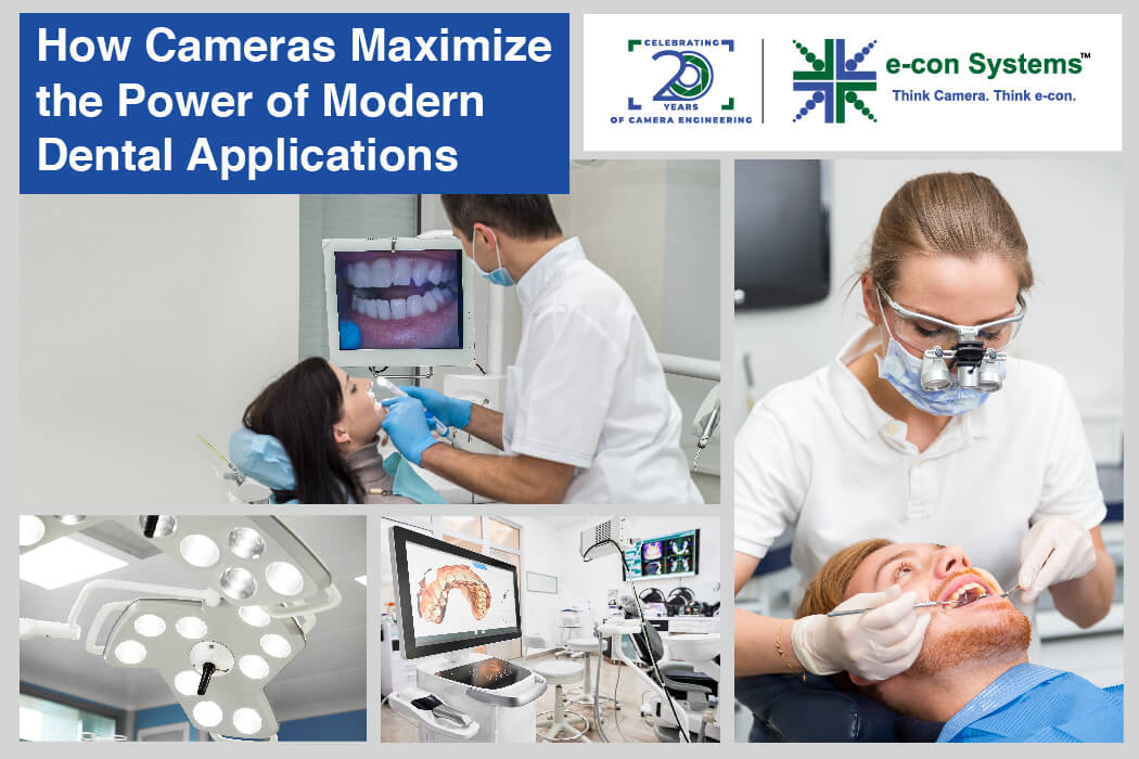
The use of cameras has changed how dental professionals diagnose, treat, and communicate with their patients. For instance, high-quality intraoral cameras that capture detailed images of teeth and gums. There are also cameras integrated into dental loupes and surgical lights to enhance critical dental processes. Other use cases include diagnostic imaging, treatment planning, etc.
The cameras provide magnified views of the oral cavity to empower dentists to perform complicated procedures with greater efficiency. Along the way, this also improves patient understanding and engagement while helping build ongoing dental care records.
In this blog, we will look at the various types of dental cameras and get insights into their key features.
Types of Cameras Used In Dentistry
Dental intraoral cameras
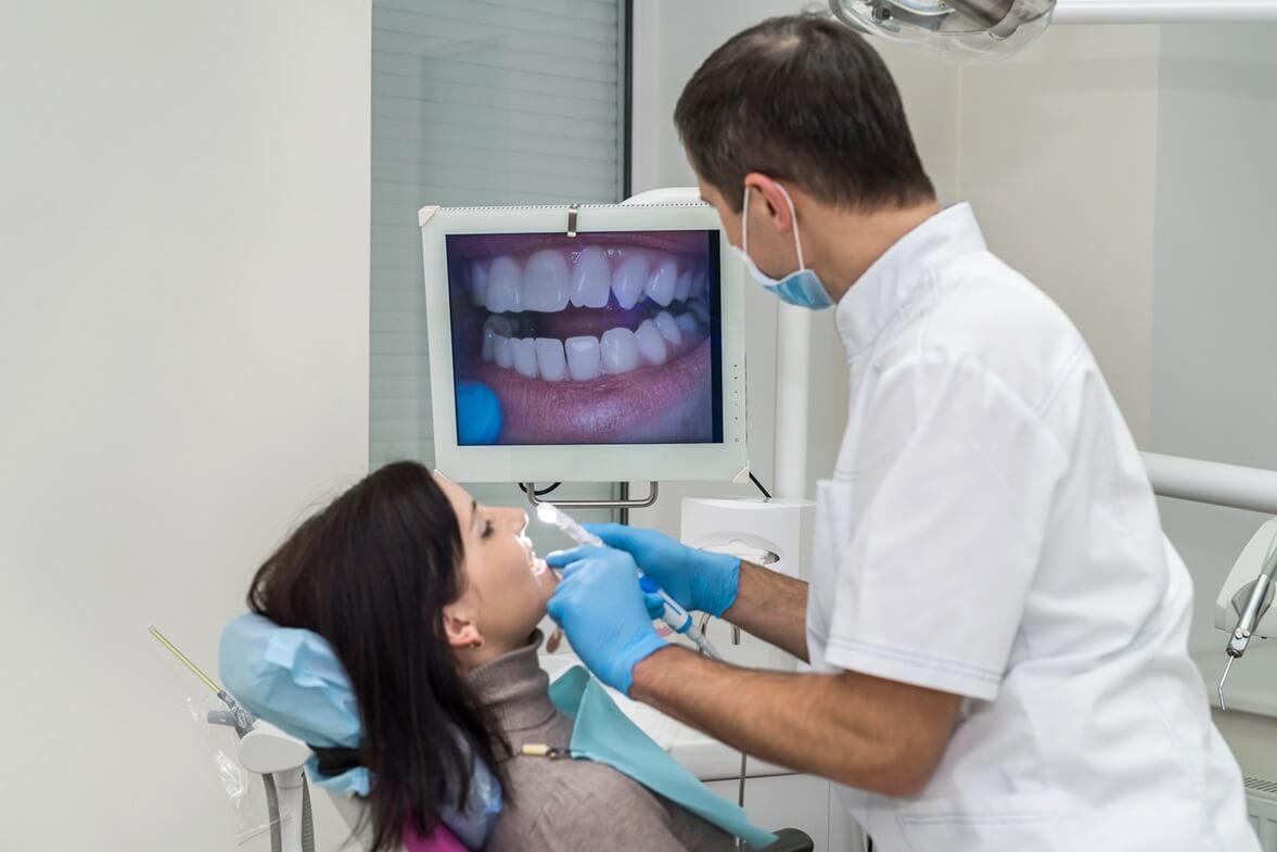
Dental intraoral cameras capture high-quality images of the insides of the oral cavity including gums, mouth, and teeth. These cameras imitate the functionalities of regular digital cameras. They also feature built-in LED lights around the lens systems to illuminate the regions of interest – as a sensor captures the images.
The compact and sleek design ensures maximum convenience for dentists.
 Dental intraoral cameras are connected to a video streaming device for real-time viewing. This helps doctors and patients view the concerned dental regions on a display monitor, making it easier to access the hard-to-reach areas of the mouth.
Intraoral scanner cameras
The cameras in intraoral scanners obtain 3-D images of the oral regions of concern to create a comprehensive view. They are used in restorative dentistry to create digital impressions for crowns, bridges, and veneers, for planning and placement of dental implants, and creating full and partial dentures.
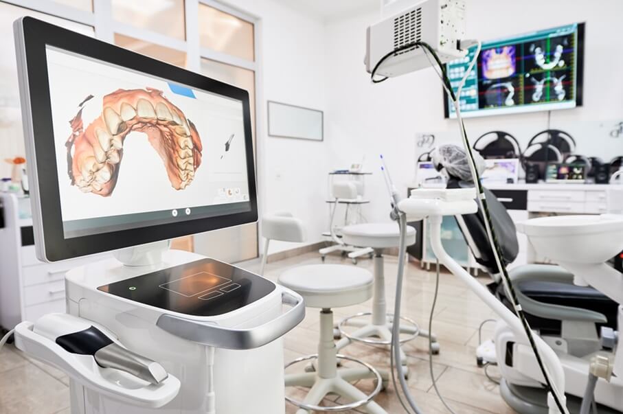
The three common imaging technologies used in intraoral scanners are:
- Confocal microscopy: A laser light is used to focus on the oral region, scanning it point by point. This data is transmitted to the camera, which acts as the detector, via a pinhole. The pinhole only allows the light focused on the specific target to reach the camera, so the camera is able to capture detailed images of the targeted oral regions.
- Structured light: A 3D scanner beams down accurate patterns of white or blue light upon the desired regions. The pattern of light gets distorted as it falls across any bends or curves in the oral area. The cameras in these scanners capture distorted light patterns frame by frame. The frames are collated to form the required 3-D image.
- Stereo vision: Stereo vision uses two cameras to capture images. The cameras are separated by a distance known as the baseline. It is important to accurately identify the same pixel in both images, known as the point of correspondence. The disparities between the two images are analyzed to achieve superior depth perception.
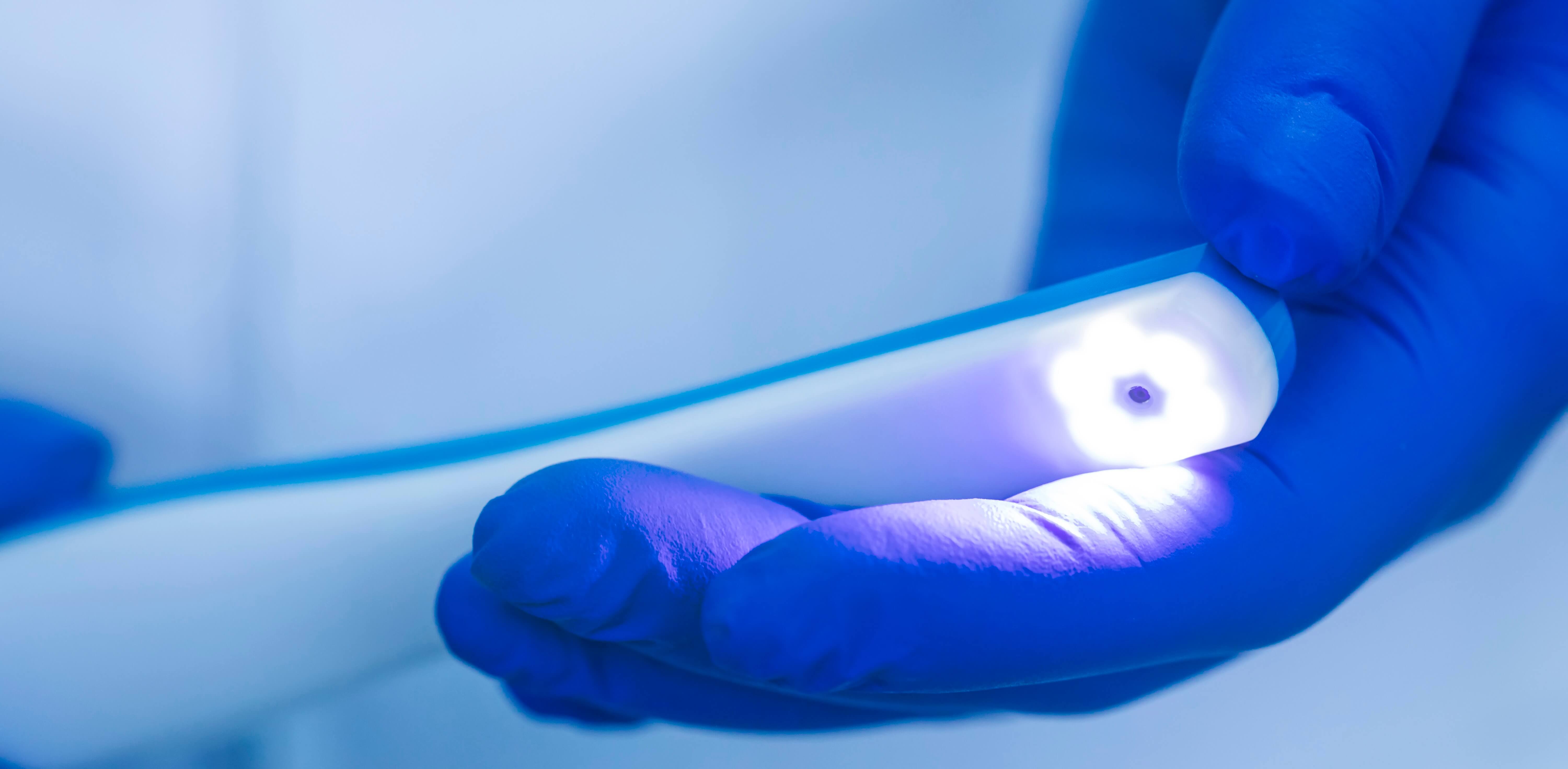
Dental loupe cameras
Dental loupes help dental professionals magnify the oral cavity, which means cameras must provide accurate streaming of their POV during complicated dental procedures. It enables other dental surgeons to visualize the dental issues at hand and the remote viewing of dental surgeries.
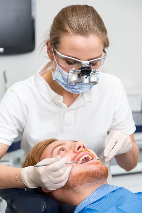
In most cases, the cameras are mounted on the frame of the dental loupes or the headband. This placement ensures that the Field of View aligns with the dentist’s line of sight. Dental loupe cameras usually come in a wearable format for a dentist’s ease of usage. They often include adjustable features to customize the fit and angle of the lenses, reducing strain on the dentist’s eyes and neck.
The magnification lenses of the dental loupe cameras, positioned in front of the dentist’s eyes, are calibrated to specific magnification levels. It typically ranges from 2.5X to 6X. Dental loupes also come with built-in LED lights to illuminate the oral cavity.
Dental surgery light cameras
Dental surgical light systems, integrated with cameras, deliver optimal illumination and capture high-resolution images and videos of procedures. They assist dental surgeons, acting as their “eyes” during surgical procedures or for remote consultation purposes.
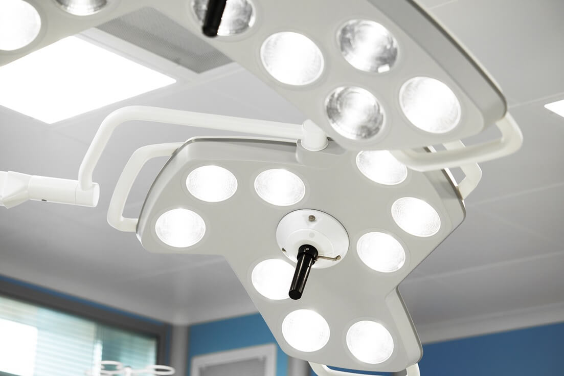
The cameras are placed near the surgical lights, which act as the ring light for illumination. These lights can be adjusted for intensity and direction as per the surgeon’s needs. However, the bright lights often cause glare or washout in the images.
Hence, the cameras used in dental surgical lights must be designed to function optimally under brightly lit conditions. The form factor of the cameras also should be suitable to fit in carious dental surgical light systems.
These cameras can also capture and record images and videos of dental surgical procedures for insurance claims and other documentation purposes.
Other Emerging Dental Imaging Solutions
- CAD/CAM imaging solutions: The use of CAD/CAM solutions is increasing in the making of zirconia crowns to achieve a better fit and inter-proximal contact. Cameras are used to obtain clinically accurate digital impressions of the patient’s oral region.
- Fluorescence/NIR imaging solutions: The method of using fluorescent/NIR light to detect dental caries has been gaining popularity in today’s world of dentistry. The lights used create contrasting image fields distinguishing dental caries from the unaffected regions. Cameras can be of great aid in the visualization of this technique.
- VR and AR imaging solutions: Virtual Reality (VR) and Augmented Reality (AR) systems are increasingly deployed in dentistry to perform complicated procedures. Currently, though, their main purpose to act as an educational tool. So, these systems require high-performance cameras to assist in creating AR/VR simulations in real time.
Dental Camera Solutions offered by e-con Systems
e-con Systems, with 20+ years of experience in designing, developing, and manufacturing OEM cameras, offers a wide range of imaging solutions to build cutting-edge dental devices. Our USB 3.0 cameras come with the best CMOS image sensors and best-fit features. They are perfect for medical use cases such as dentistry, point of care, fundus imaging, remote patient monitoring, surgery, etc.
Explore all our dentistry cameras
Check out the Camera Selector Page to see our full portfolio.
If you want to get started integrating our cameras into your dental products, please write to camerasolutions@e-consystems.com.

Balaji is a camera expert with 18+ years of experience in embedded product design, camera solutions, and product development. In e-con Systems, he has built numerous camera solutions in the field of ophthalmology, laboratory equipment, dentistry, assistive technology, dermatology, and more. He has played an integral part in helping many customers build their products by integrating the right vision technology into them.