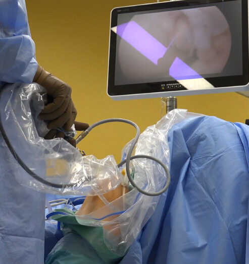
Surgical visualization and imaging systems are an integral part of modern surgeries, especially tumor detection. They provide surgeons with real-time, high-definition views of internal structures for enhanced accuracy and safety. These technologies are crucial for minimizing damage to healthy tissues, improving accuracy in minimally invasive procedures, and offering real-time feedback.
Multi-modal imaging is one of the common components of surgical visualization systems. It refers to the use of multiple imaging techniques simultaneously or sequentially to gain a thorough understanding of a subject. In the medical field, multi-modal imaging combines data from different sources, such as RGB (visible light), infrared (IR), ultrasound, PET, or fluorescence imaging, into a single cohesive framework. It improves the diagnostic and therapeutic process by providing complementary information from each modality.
RGB-IR cameras, which capture both visible and infrared light, offer a distinctive advantage. In this blog, you’ll discover how these cameras can simultaneously enhance the visualization of surface details through RGB imaging and subsurface structures using IR imaging. You’ll also get expert insights on how RGB-IR cameras are transforming medical and surgical visualization.
How Do RGB-IR Cameras Work?
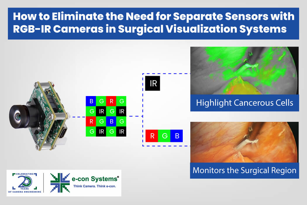
RGB-IR cameras are equipped with sensors featuring dedicated RGB and IR pixels, unlike ordinary sensors with a Bayer BGGR pattern. The unique Color Filter Array (CFA) in RGB-IR cameras includes pixels that exclusively allow IR light, enabling separate RGB and IR data output without mixing, which is crucial for medical applications like fluorescence-guided surgery. Some of their benefits are:
- High imaging performance in all lighting conditions
- Compact design that eliminates the need for dual cameras
- Longer camera and device lifetimes due to non-mechanical switching
- Intelligent color correction through a dedicated IR channel
When designing an embedded RGB-IR camera, key components must be tailored for this technology. The sensor must support a pixel array with both RGB and IR pixels, with manufacturers like onsemi and OmniVision offering suitable options. Optics require a dual bandpass filter lens to allow both visible and NIR light, replacing conventional lenses with IR-cut filters. Finally, the Image Signal Processor (ISP) plays a critical role in separating RGB and IR frames and correcting IR contamination on RGB channels, which requires advanced algorithms and tuning expertise.
How RGB-IR Cameras Enhance Medical and Surgical Visualization
Image-Guided Surgeries
Image-Guided Surgeries (IGS) use preoperative or intraoperative images to directly or indirectly guide surgeons during operations. These systems provide detailed views of the surgical area for precise navigation while reducing risks to surrounding tissues. IGS systems are valuable for performing complex procedures like neurosurgery, orthopedic surgery, and minimally invasive interventions.
Such surgeries are increasingly incorporated into robotic surgery and AR-guided surgical systems. Among the various image-guided surgery techniques, fluorescence-guided surgery is a widely used method.

Fluorescence-guided surgeries use fluorescent dyes to visualize and differentiate tissues. The dyes, when exposed to specific wavelengths of light, emit fluorescence that special imaging systems can capture. It enables surgeons to see structures that are otherwise invisible under normal lighting conditions.
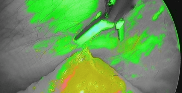
Indocyanine Green (ICG) is one of the most common fluorophores used in human surgeries due to its wide margin of safety. ICG is a Near-Infrared (NIR) dye that binds to proteins in the blood and fluoresces a bright green color when illuminated by NIR light.
Fluorescence-guided surgeries typically occur in two ways:
- Open surgery: The surgeon directly accesses the surgical site through an incision. Fluorescent dyes, such as ICG or 5-ALA, are administered to the patient, and the surgical area is illuminated with a specific wavelength of light that excites the dye. It causes the dye to fluoresce, highlighting specific tissues, such as tumors, blood vessels, or lymph nodes. Hence, it provides a broader view of the surgical field and a more direct hands-on control of tissues.
- Endoscopic Surgery: The surgery is performed using a minimally invasive approach. A rigid endoscope, a solid and inflexible tube equipped with a camera and light source at the tip, is inserted through small incisions or natural body openings. Then, the fluorescent dye is injected into the patient as the endoscope’s light source excites the dye, causing the targeted tissues to fluoresce. It is used in procedures like sentinel lymph node mapping, gastrointestinal surgeries, and laparoscopic cancer resections, where minimizing tissue damage and recovery time is important.
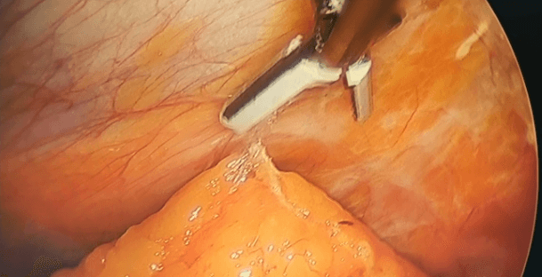
In both methods, RGB imaging offers a clear view of the surgical field while IR imaging highlights the fluorescent dye. This enables accurate identification of critical structures like blood flow, tumor margins, and lymphatic pathways. Ultimately, it enhances the surgeon’s ability to differentiate tissues, improving the accuracy and safety of the procedure.
Cancer detection and diagnosis
Tumor cells exhibit distinct properties such as altered optical characteristics, increased metabolic activity, and enhanced vascularization. They often absorb, and scatter light differently compared to normal tissues due to variations in tissue density, blood supply, and cellular structure.
Tumors also tend to have higher metabolic rates, leading to changes in oxygenation levels and localized temperature differences, which can be detected. The process of angiogenesis, where tumors develop their own blood vessels, distinguishes them from healthy tissues, making them more visible in the type of imaging that can capture these variations.


Therefore, RGB-IR cameras play a crucial role in tumor detection by leveraging their ability to capture visible light (RGB) and infrared (IR) light. The RGB component provides clear visualization of tissue structures, including color differentiation, while the IR component enhances imaging by penetrating deeper into tissues and revealing details such as variations in tissue composition, vascular structures, and oxygenation levels.
That’s how RGB-IR cameras offer improved contrast and functional insights, making it easier to differentiate between healthy and malignant tissues.
What’s New with e-con Systems’ RGB-IR Cameras?
Since 2003, e-con Systems has been designing, developing, and manufacturing OEM cameras. We are thrilled to announce the launch of See3CAM_CU83, our latest 4K AR0830 RGB-IR USB 3.2 Gen 1 camera for applications that require simultaneous visible and IR imaging. With its perfect combo of sensor, optics, and ISP, we are confident that it can address several concerns faced by medical and life sciences’ companies.
Our proven track record in advanced customization also ensures that you no longer have to go through long design lifecycles to design and deploy your high-end MedTech applications.
Check out our Medical Market Page to know more about our imaging expertise for medical applications,
You can also use our Camera Selector to get a full view of our camera portfolio.
If you need any help in picking and integrating the right cameras into your medical devices, please write to camerasolutions@e-consystems.com.

Balaji is a camera expert with 18+ years of experience in embedded product design, camera solutions, and product development. In e-con Systems, he has built numerous camera solutions in the field of ophthalmology, laboratory equipment, dentistry, assistive technology, dermatology, and more. He has played an integral part in helping many customers build their products by integrating the right vision technology into them.


